Importance of the cuticular layer during the colonization of the fungus that causes negrilla in Agave salmiana Otto ex Salm-Dyck ssp. salmiana
DOI:
https://doi.org/10.29298/rmcf.v13i70.1265Abstract
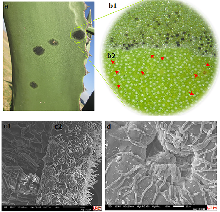
The Agave genus plants represent a non-timber forest resource, valuable for soil recovery. The epidermis of the agave leaves contains multiple stomata and is covered by a cuticular layer. Currently, agave plants have a fungal disease that is characterized by circular gray spots on the maguey leaves, which over time become necrotic and eventually, these lesions end up drying them. The aim of this work was to describe the cuticular layer during the fungus colonization that causes bold in Agave salmiana subsp. salmiana. The cuticular layer is 121 mm ± 2.8 thick. A homogeneous distribution of the stomata was observed, and the density (22.67-27.67 stomata mm-2) and the stomatal index (10.61-14.15) were determined. Tetracytic stomata and isodiametric polygonal epidermal cells were identified, the ostioles size were calculated as 57.9 mm ± 5 long and 23.75 mm ± 1.25 wide. The transverse and paradermal sections showed that the fungal hyphae and appressoria are restricted from the obverse side of the cuticular layer, which confirms the importance of preserving the epidermis in the maguey pulquero leaves.
Downloads
References
Bernardino-Ninacor, A., R. Mora-Escobedo, J. L. Montañez-Soto, S. Filardo-Kerstupp and L. González-Cruz. 2012. Microstructural differences in Agave atrovirens Karw leaves and pine by age effect. African Journal of Agricultural Research 7(24):3550-3559. Doi:10.5897/ AJAR11.1185. DOI: https://doi.org/10.5897/AJAR11.1185
Biswapriya, B. M., R. A. Biswa, D. Granot, S. M. Assmann and S. Chen. 2015. The guard cell metabolome: functions in stomatal movement and global food security. Frontiers in Plant Science 6:1-13. Doi:10.3389/fpls.2015.00334. DOI: https://doi.org/10.3389/fpls.2015.00334
Bury, M., A. Andolfi, B. Rogister, A. Cimmino, V. Mégalizzi, V. Mathieu, O. Feron, A. Evidente and R. Kiss. 2013. Fusicoccin A, a phytotoxic carbotricyclic diterpene glucoside of fungal origin, reduces proliferation and invasion of glioblastoma cells by targeting multiple tyrosine kinases. Translational Oncology 6(2):112-23. Doi: 10.1593/tlo.12409. DOI: https://doi.org/10.1593/tlo.12409
Comité Estatal de Sanidad Vegetal de Guanajuato A.C. (CESAVEG). 2008. Manual de plagas y enfermedades del Agave. Casa editorial. Irapuato, Gto., México. 28p.
Chávez-Güitrón, L. E., F. C. Salinas-Pérez, E. A. Pérez-Salinas, J. Caballero, A. Vallejo-Zamora y E. Sandoval-Zapotitla. 2019. Variación de caracteres epidérmico-foliares de Agave salmiana subsp.salmiana (Asparagaceae) en el centro de México. Botanical Sciences 97 (4): 711-724. Doi: 10.17129/botsci.2159. DOI: https://doi.org/10.17129/botsci.2159
Y
García M., A. 2007. Los agaves de México. Ciencias 87:14-23. https://www.redalyc.org/pdf/644/64408704.pdf (20 de enero de 2021).
Gentry, H. S. and J. R. Sauck. 1978. The stomatal complex in Agave: groups Deserticolae, Campaniflorae, Umbelliflorae. Proceedings of the California Academy of Sciences series 41:371–387. https://ia800206.us.archive.org/0/items/biostor-78394/biostor-78394.pdf (25 de octubre de 2020).
Gudesblat, E. G., S. P. Torres and A. A. Vojnov. 2009. Stomata and pathogens: warfare at the gates. Plant Signaling & Behavior 4(12):1114-1116. Doi: 10.4161/psb.4.12.10062. DOI: https://doi.org/10.4161/psb.4.12.10062
Hernández-Valencia, R. E. M., R. López-Franco and A. Benavides-Mendoza. 2003. Micromorfología de la epidermis foliar de Agave tequilana Weber. Agrofaz 3(2):387-396. https://www.researchgate.net/publication/283995003 (15 de octubre de 2020).
Lee S., H. Choi, S. Suh, I.S. Doo, K. Y. Oh, E. J. Choi, A. T. S. Taylor, P. S. Low and Y. Lee. 1999. Oligogalacturonic acid and chitosan reduce stomatal aperture by inducing the evolution of reactive oxygen species from guard cells of tomato and Commelina communis. Plant Physiology 121:147-52. Doi:10.1104/pp.121.1.147. DOI: https://doi.org/10.1104/pp.121.1.147
Lee, J. S. 2010. Stomatal opening mechanism of CAM plants. Journal of Plant Biology 53:19–23. Doi:10.1007/s12374-010-9097-8. DOI: https://doi.org/10.1007/s12374-010-9097-8
Mendgen, K., M. Hahn and H. Deising. 1996. Morphogenesis and mechanisms of penetration by plant pathogenic fungi. Annual Review of Phytopathology 34:367–86. Doi: 10.1146/annurev.phyto.34.1.367. DOI: https://doi.org/10.1146/annurev.phyto.34.1.367
Neto, I. L. C. and F. M. Martins. 2012. Anatomia dos órgãos vegetativos de Agave sisalana Perrine ex Engelm (Agavaceae). Revista Caatinga 25(2):72-78. https://www.redalyc.org/pdf/2371/237123825011.pdf (12 de febrero de 2021).
Nobel, P.S. 1994. Remarkable Agaves and Cacti. Cambridge University Press. New York, NY, USA. 166 p.
Pérez-España, V. H., J. A. Cuervo-Parra, C. Paz-Camacho, M. A. Morales-Ovando, C. A. Gómez-Aldapa, G. C. Rodríguez-Jimenes, V. J. Robles-Olvera, P. A. López-Pérez and T. Romero-Cortés. 2019. General characterization of cuticular membrane isolated from Agave salmiana. International Journal of Bio-resource and Stress Management 10(1):046-052. Doi:10.23910/IJBSM/2019.10.1.1950. DOI: https://doi.org/10.23910/IJBSM/2019.10.1.1950
Reina-Pinto, J. J. and A. Yephremov. 2009. Surface lipids and plant defenses. Plant Physiology and Biochemistry 47(6):540-549. Doi:10.1016/j.plaphy.2009.01.004. DOI: https://doi.org/10.1016/j.plaphy.2009.01.004
Roth I., T. Merida y H. Lindorf. 1986. Morfología y anatomía foliar de plantas de la Selva Nublada de Rancho Grande. Parque Nacional “Henry Pittier”. El ambiente físico, ecología general y anatomía vegetal. Fondo Editorial Acta Científica Venezolana. Caracas, Venezuela. pp. 205-241.
Sosa, C. M., G. S. Alemán, H. Y. Pérez, C. E. Abreu, C. D. Sosa and O. G. González. 2014. Caracterización de la lámina foliar de plantas de Agave fourcroydes Lem. obtenidas por propagación asexual. Biotecnología Vegetal 14(1):37-44. https://revista.ibp.co.cu/index.php/BV/article/view/40/433 (12 de febrero de 2021).
Staiger, S., P. Seufert, K. Arand, M. Burghardt, C. Popp and M. Riederer. 2019. The permeation barrier of plant cuticles: uptake of active ingredients is limited by very long-chain aliphatic rather than cyclic wax compounds. Pest Management Science 75(12):3405-3412. Doi: 10.1002/ps.5589. DOI: https://doi.org/10.1002/ps.5589
Staples, R. C. and V. Macko. 1980. Formation of infection structures as a recognition response in fungi. Experimental Mycology 4(1):1-15. Doi:10.1016/0147-5975(80)90045-6. DOI: https://doi.org/10.1016/0147-5975(80)90045-6
Tafolla-Arellano, J. C., A. González-León, M. E. Tiznado-Hernández, L. Zacarías G. and R. Báez-Sañudo. 2013. Composición, fisiología y biosíntesis de la cutícula en plantas. Revista Fitotecnia Mexicana 36(1):3-12. http://www.scielo.org.mx/scielo.php?script=sci_arttext&pid=S0187-73802013000100001&lng=es&tlng=es (20 de noviembre de 2020). DOI: https://doi.org/10.35196/rfm.2013.1.3
Vargas-Rodríguez, L., M. I. García-Vieyra, B. I. León-Bata and P. Lozano-Sotomayor. 2017. Physical properties and microscopic structure of the Agave salmiana cuticle (mixiote). Revista Chapingo Serie Zonas Áridas 17(2):1-9. Doi: 10.5154/r.rchsza.2017.12.017. DOI: https://doi.org/10.5154/r.rchsza.2017.12.017
Wilkinson, H. 1979. The plant surface (mainly leaf). In: C. R. Metcalfe and L. Chalk (eds.). Anatomy of Dicotiledons. Oxford Claredous Press. London, UK. pp. 97-16.
Zeng, W. and S. Y. He. 2010. A prominent role of the flagellin receptor FLAGELLIN-SENSING2 in mediating stomatal response to Pseudomonas syringae pv tomato DC3000 in Arabidopsis. Plant Physiology 153:1188–1198. Doi: 10.1104/pp.110.157016. DOI: https://doi.org/10.1104/pp.110.157016
Published
How to Cite
Issue
Section
License

This work is licensed under a Creative Commons Attribution-NonCommercial 4.0 International License.
The authors who publish in Revista Mexicana de Ciencias Forestales accept the following conditions:
In accordance with copyright laws, Revista Mexicana de Ciencias Forestales recognizes and respects the authors’ moral right and ownership of property rights which will be transferred to the journal for dissemination in open access.
All the texts published by Revista Mexicana de Ciencias Forestales –with no exception– are distributed under a Creative Commons License Attribution-NonCommercial 4.0 International (CC BY-NC 4.0), which allows third parties to use the publication as long as the work’s authorship and its first publication in this journal are mentioned
The author(s) can enter into independent and additional contractual agreements for the nonexclusive distribution of the version of the article published in Revista Mexicana de Ciencias Forestales (for example, include it into an institutional repository or publish it in a book) as long as it is clearly and explicitly indicated that the work was published for the first time in Revista Mexicana de Ciencias Forestales.
For all the above, the authors shall send the form of Letter-transfer of Property Rights for the first publication duly filled in and signed by the author(s). This form must be sent as a PDF file to: ciencia.forestal2@inifap.gob.mx
This work is licensed under a Creative Commons Attribution-Noncommercial 4.0 International license.